Teesside University researchers are using artificial intelligence (AI) to enhance cancer diagnostics, training deep learning models to improve accuracy and revolutionise treatment.
Their work aligns with a global shift towards AI-powered healthcare solutions, where enhanced diagnostics and treatment precision are improving patient outcomes and optimising healthcare efficiency.
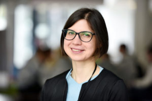
In this guest blog, Professor Annalisa Occhipinti, explains how use of AI and light could improve cancer diagnosis. It also coincides with the UK’s Breast Cancer Awareness Month during October, when people and organisations work together to highlight the importance of cancer awareness, education and research.
Professor Occhipinti leads the AI group in the University’s School of Computing, Engineering & Digital Technologies.
When diagnosing cancer, a crucial step involves examining a small tissue sample under a microscope.
This is called a biopsy, and the detailed images taken from it help doctors decide whether the cells are normal or cancerous.
These images are known as histopathological images – high-resolution pictures of how tissue looks under the microscope.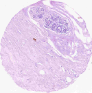
The microscope image above shows breast tissue treated to highlight the structure of cells. This type of image helps doctors to see the detailed arrangement and shape of cells within the tissue, which is important for identifying healthy areas and detecting potential abnormalities like cancer.
These images are incredibly useful, as they show the shape of the cells, how they are arranged, and whether anything looks abnormal. However, interpreting them is not always easy. It takes years of training, and even then, small differences between healthy and cancerous cells can sometimes be hard to spot. That is where AI can help.
New developments
In the University’s School of Computing, Engineering & Digital Technologies, academic researchers are developing a new approach that combines AI with two types of information:
- Microscope images of tissue samples (histopathological images)
- Infrared spectroscopy, a method that shines light on the tissue to find out what it is made of at a chemical level.
While the microscope images show what the tissue looks like, infrared spectroscopy tells us what the tissue is made of. By combining both, we can get a much more complete picture.
How does it work?
We use AI to bring all this information together. In particular, the AI model is trained to recognise patterns from both the images and the chemical data. And it learns how to classify healthy tissue or cancerous tissue more accurately than if we used just one type of data alone.
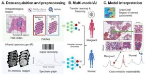
The diagram above shows the step-by-step process of our AI method. Starting with tissue samples, we capture microscope images to analyse the shape of cells and use infrared spectroscopy to analyse the chemical structure of cells. Then, we train an AI model to combine both images and classify the tissue as either cancerous (malignant) or healthy (normal).
Why This Matters?
This combined approach could lead to more accurate and earlier cancer diagnoses. It can reduce uncertainty, support pathologists in making decisions, and help doctors choose the best treatment sooner.
In the future, this technology could be used in hospitals to make cancer diagnosis not only more precise but also more consistent, ensuring that every patient gets the best possible care from the very beginning.
If you would like to know more about this project or other AI projects developed in the group, contact Professor Occhipinti on a.occhipinti@tees.ac.uk

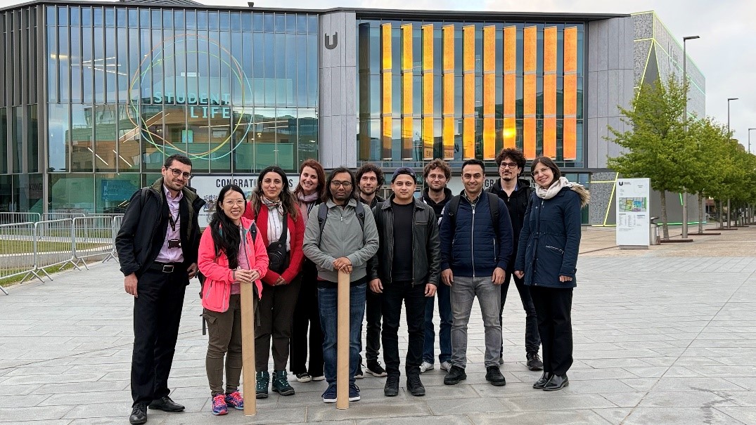
Members of the Teesside University team involved in the research project, which was funded by the CLIRPath-AI Network, EPSRC Funding, are pictured above. Also part of the research team was Teesside University lecturer Aliyu Abubakar.
Find out more about Professor Occhipinti