Lecturer in Environmental Science (2 Posts)
Our Department is advertising for two permanent positions of lecturer in environmental science. The newly appointed lecturers have an opportunity to take a leading role in our Earth, Ecology and Environment research collective and bring their own research and/or consultancy expertise.
The job ad can be found following the two links below:
Job.ac.uk
If you would like to discuss how your research could fit within the Earth, Ecology and Environment research collective – please get in touch with Ambroise a.baker@tees.ac.uk.
Slogging for Skelton
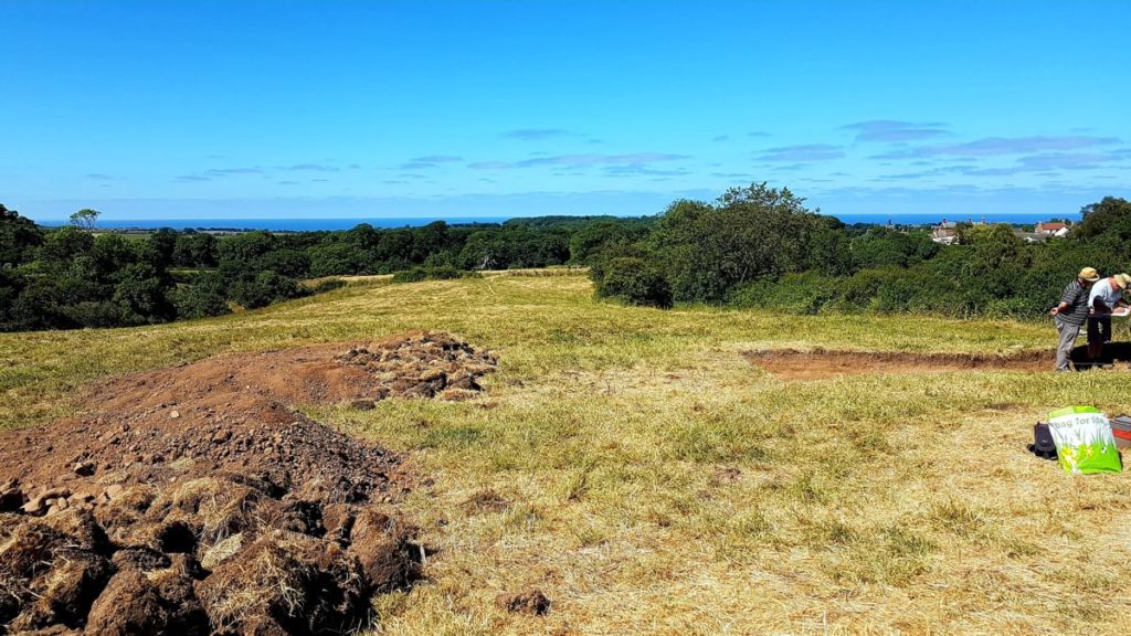
Just this week, our new paper on “Mapping an archaeological site: Interpreting portable X-ray fluorescence (pXRF) soil analysis at Boroughgate, Skelton, UK” was published! And so, we thought it would be nice to share some of the work that went toward this with you all.
Boroughgate was a 12th Century medieval borough in Skelton, North Yorkshire UK, near the All Saint’s Old Church and Skelton Castle. It was placed in the perfect location to support trade and income for the castle via but unfortunately it was unsuccessful and abandoned around 1400 CE. The remnants of earthworks at the site and medieval documentation recording some of the tradespersons at Boroughgate gave some clues as to the history of the site. Tees Archaeology went through a series of surveys before excavating the site, inviting us out to complete some pXRF analysis and explore whether pXRF elemental analysis can enhance and support their interpretations of the site. This was also an excellent opportunity for us to show the value of our method development for pXRF soil analysis in archaeology! Admittedly, this also may have been a bit of an excuse to get out on such a glorious Summers day…
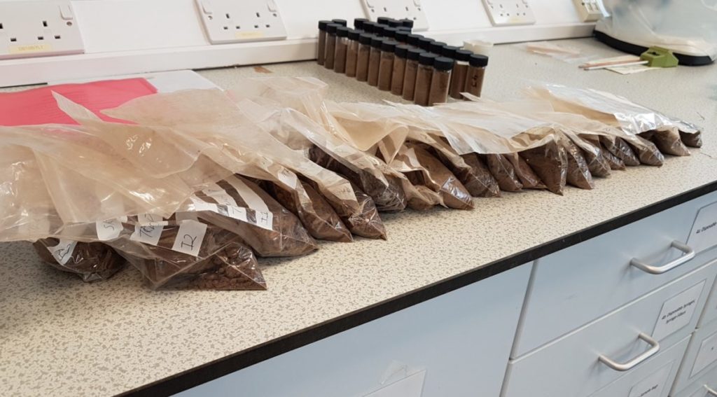
pXRF is often seen as a rapid point-and-shoot method but for good quality data, we really need an appropriate methodology. The soil matrix can vary greatly over just short distances, and we need to make sure that all our soil is examined in the same way, otherwise our comparisons are inconsistent and not well validated. We extract soil samples, dry them in the lab (preferably oven dried), grind down and sieve the samples so they’re nice and homogenous, and prepare them into pXRF sample cups. This does of course mean that we end up with a fair bit of soil samples in the lab from just one small area of soil..!
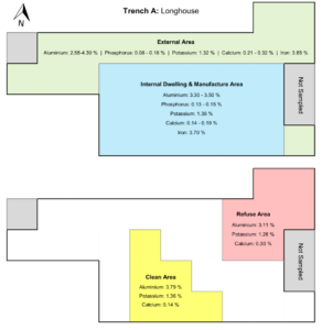
Research into social organisation and the activities or use of space from archaeological excavations uncover hidden knowledge on past societal practices and the structuring of historic communities. This work explored whether we could map out the elemental distribution of soil to identify different activity areas. This is discussed in much more detail in the journal article but just briefly, the distribution of aluminium, phosphorus, potassium calcium and iron distinguished between the internal dwelling and external area of a longhouse. Aluminium, potassium and calcium also distinguished a likely clean or food preparation area against a refuse area. These areas also aligned closely with the locations of artefacts such as pottery fragments, daub, and domestic or charred waste, as well as structural remains such as building foundation pads, postholes and wall foundations.
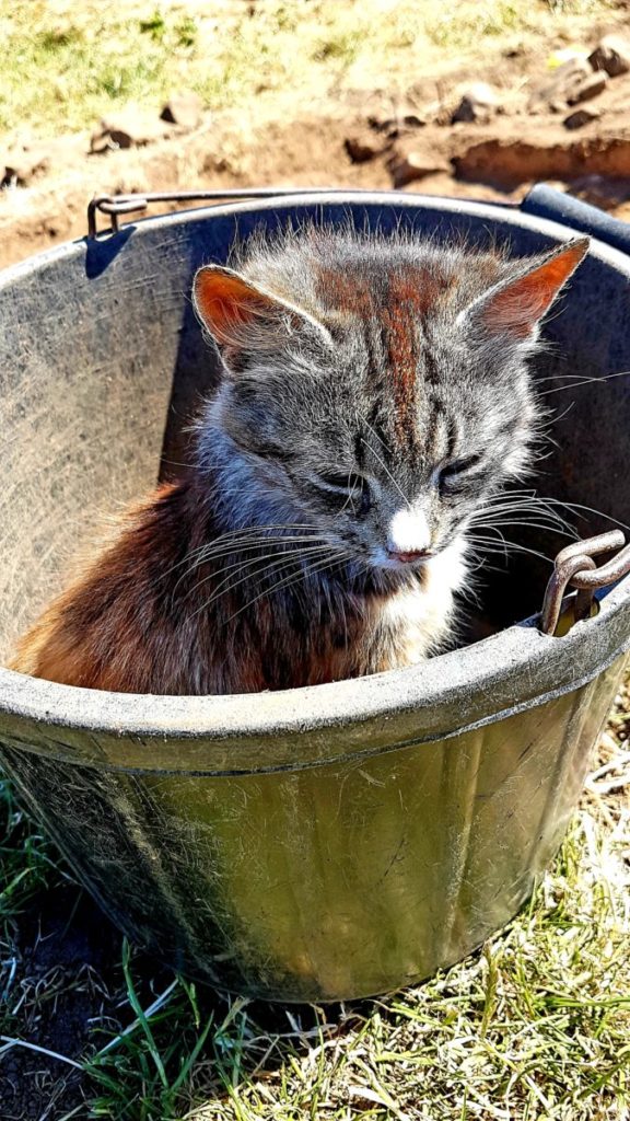
This was a well worthwhile investigation into mapping pXRF of soil which we’re very excited to continue further. Don’t hesitate to contact us if you’re interested in surveying your site with pXRF, we’d love to see how much more we can learn about past communities with pXRF! Now before you go, don’t forget to say hi to the ridiculously sweet kitten which I’ve dubbed Sandy the Archaeology Cat because of its love for sitting in soil buckets and climbing over your shoulders judging your use of the Harris Matrix:
And finally, thanks to David Errickson at Cranfield University, and Tees Archaeology for inviting us out to your site!
TUBA
How can microbes can turn rubbish into riches?
Our own Dr Caroline Orr took part to the Royal Society’s Summer of Science 2021.
An interactive online game was also created:
https://royalsociety.org/summer-science/summer-science-2021/urban-landscape/summer-microbes
Research grant to study geochemical and microbial conditions underpinning turf preservation at the Roman site of Vindolanda, UK
Drs G Taylor and C Orr won a new grant to carry out the work as follows.
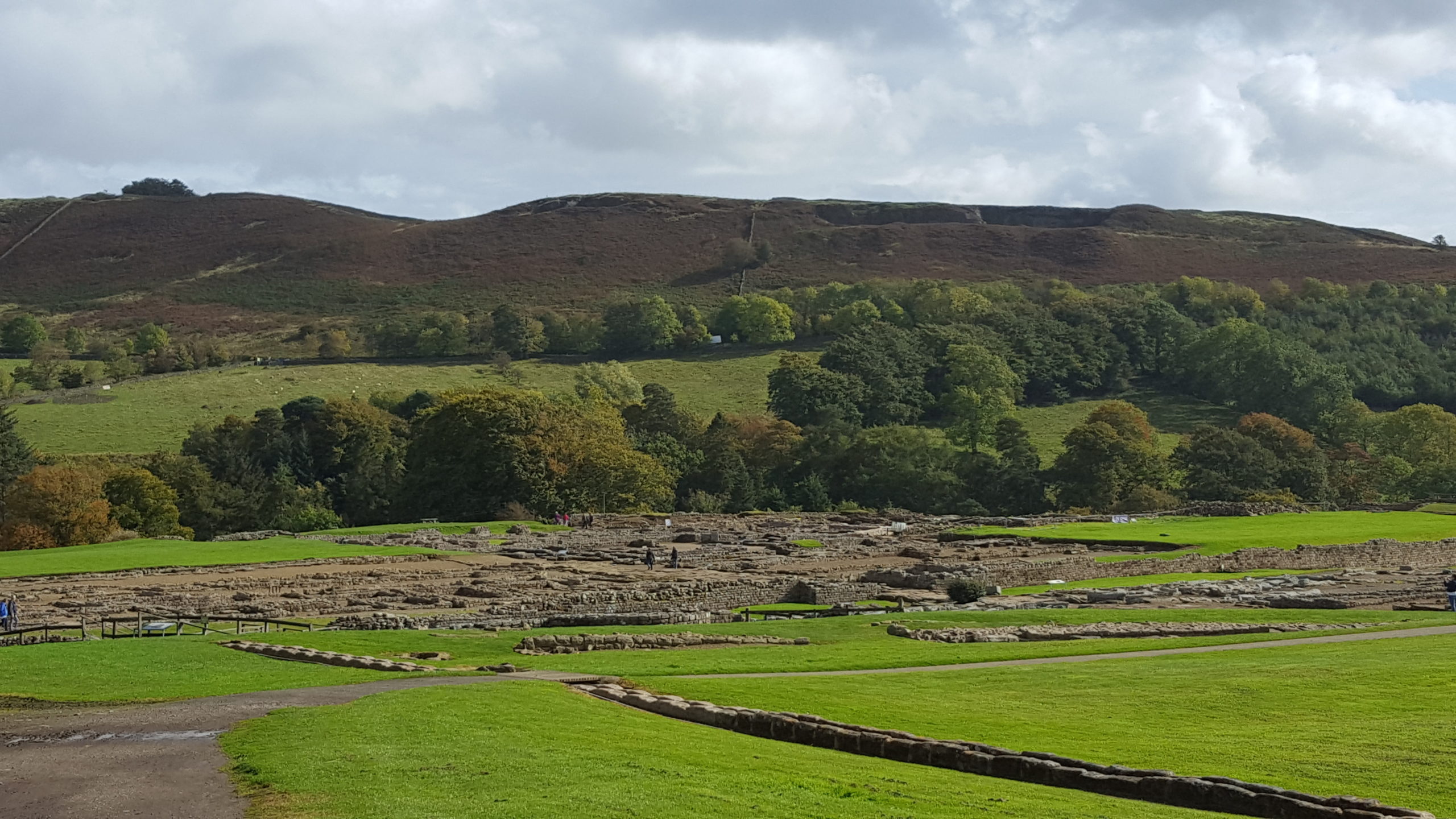
The Roman Fort site at Vindolanda is known for the exceptional preservation of its finds, among them wooden writing tablets and leather shoes. A recent study into Roman construction practices at the site demonstrated that this preservation extends to the turf ramparts, with plant fibres and seed heads still visible and microbes seemingly surviving within the soils. While that earlier project focused on turf in building, this new one will evaluate what this same material can reveal about the ancient environment. As a pilot study, it will assess the geochemical and microbial conditions, which underpin this preservation, and evaluate the turf blocks’ potential as environmental archives to reconstruct the landscape around the fort through time. Results will inform four smaller-scale follow-on analytical packages and three large-scale interdisciplinary funding applications to investigate the economic and ecological impacts of turf sourcing and turf’s potential as a zero-carbon building material for the future
Sustainable Drainage Research at Climate Exp0
Dr Ed Rollason, along with colleagues at Durham University and University College Dublin are collaborating on a project exploring how we conceptualise Sustainable Drainage Systems (SuDS) as mechanisms for enhancing high density urban environments. They are currently exhibiting a poster on the work at Climate Exp0, a free conference being run as a prequel to the COP26 climate summit to be held later in the year.
Sustainable drainage systems are key components of urban drainage infrastructure for new build houses. However, retrofit takeup of SuDS is low and generally unimaginative, and projects often do not meet their aspirations for delivering multiple benefits. We argue that identifying the effectiveness and potential for retrofitting SuDS requires understanding the nexus between the nature of the problem being addressed, the place in which the intervention is being implemented, and the level of investment which is being made available. This paper will propose a new conceptual model integrating these factors which will allow SuDS designers and promoters to better understand where and how to implement SuDS to achieve the greatest chances of success and the greatest co-benefits.
pXRF on pathology! Catching up with a student publication
Recently, we worked with Naomi Kilburn, a Master’s student at Durham University, whose dissertation project titled ‘Assessing pathological conditions in archaeological bone using portable X-ray fluorescence (pXRF)’ was published just this month! Fab, right? We took a moment from our calendar of Teams calls to have a Zoom call with Naomi and catch up on her work, experience, and the research.

Hi Naomi! So first off, tell us about yourself – what’s your research passion?
My passion is for palaeopathology – I love looking at human skeletons to see what they can tell us about health, diseases, and life in the past.
Oh wow, fascinating! What area of palaepathology do you enjoy the most?
There are so many fascinating areas to explore, but… at the top are studying infant and childhood health and looking for ways to expand how we learn about health in the past.
What pathway did you take to get into palaeopathology?
I recently completed my master’s at Durham University and I’m currently working on securing some PhD funding so that I can keep asking (and maybe sometimes even answering) exciting questions about people and their bones.
So your paper, Assessing Pathological Conditions in Archaeological Bone using pXRF… how did that get started?
Well, this project came about through talking with Becky Gowland, my advisor at Durham, about possible dissertation projects.
Becky suggested portable X-ray fluorescence (generally called pXRF, as otherwise it’s quite a mouthful) as a way to combine studying children with a new palaeopathological technique.
My major research question was thus formed: Can pXRF be used to distinguish between different diseases in archaeological bone?
Mmm yes I can see how that idea was formed! Were you ready and raring to go or did you have a couple more hurdles to jump?
Ah yes, so, with the project idea settled, I then needed to figure out how to access a pXRF. Luckily, Becky knows many people and put me in contact with Tim Thompson at Teesside University.
After getting the go-ahead from Tim, I carefully packed some femora into boxes and headed to Teesside.
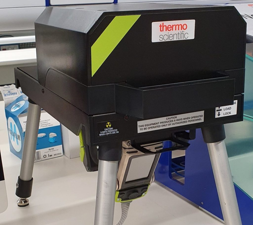
Excellent! How did you find coming to Teesside for a few days?
Rhys and Helga rolled out the welcome mat, showed me around the campus and gave me a crash course in using pXRF. And bingo, I was all set!.. until some unexpected hiccups…
Oh no! What happened?
The pXRF stopped working properly and had to be repaired, which muddles up all the project timelines. Disaster! (Okay, so it wasn’t that much of a disaster). But, with Helga’s supreme organisation and flexibility of everyone using the pXRF, things were quickly back on track better than ever!
Glad to hear it was sorted out! So… what did the pXRF do?
With pXRF, I could zap the bones with X-Rays and find out what kinds of elements are in the bones (and how much of them there is!).
What did this tell you?
I found that the real time-consuming part of pXRF was playing with all the numbers and figuring out what they might mean. My summer was spent making scatterplots and doing statistical tests to try and tease out patterns in the data that could be related to scurvy, or rickets, or any of the other diseases I was looking at.
Data, data, data! What did you find out?
The patterns remained elusive (science!), but the search was fun! I looked at elemental ratios potentially related to cribra orbitalia, neoplastic disease, rickets, scurvy, syphilis, and pathological new bone formation. Unfortunately, elemental ratios were more closely related to post-burial processes, but examining larger sample sizes of each pathology could shed light on new information.
I see! Did you find out anything else?
Actually, I found out how useful the pXRF is! This work couldn’t have been done without pXRF because it allows rapid and non-destructive analysis (can’t go chopping up and grinding down archaeological collections willy-nilly!).
Awesome, go Team pXRF!
It’s absolutely fantastic to see students get their work get published, it’s such a great boon for PhD application process. I’m sure you’ll join us in wishing Naomi all the best in her bright academic future, we look forward to seeing what comes next!
TUBA
Telling the Beavers
You may have seen recently some talk about the reintroduction of certain animals into the UK. There are a few animals you might never have realised were native to the UK, such as lynx and bears, the white-tailed eagle, and the ridiculously cute pine marten (seriously, look at them!). Well, we’ve just started work on a project alongside our Ecology and Environmental friends at Teesside for the Forestry Commission investigating a new beaver enclosure!
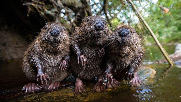
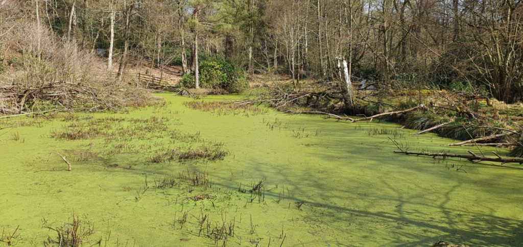
A beaver’s paradise
Within just a year, what started as a small stream passing through the private woodlands has now become home to two beavers, their four new-born kits (yeah, I wish they were called babe-eavers too) and this massive pond teeming with new aquatic life! And to think, you used to be able to stroll through here without needing overalls and a raft just a year ago…
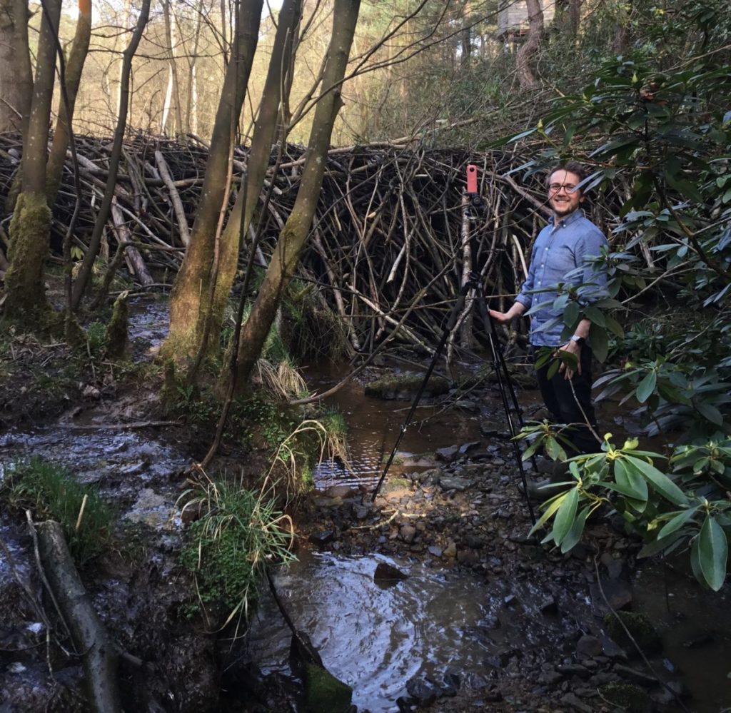
Why do we give a dam?
Beavers have a pretty well-known habit of building dams. Did you know that a major reason for this is winter survival? The deep water behind the dam doesn’t freeze the whole depth, allowing the beavers to anchor a food source at the bottom of the water and survive the winter. When building the dams, the beavers scurry around the environment selecting the juiciest of trees and have a little nibble. Okay, more like a feast. As they munch on the bark, the trees eventually give way and topple over. Sometimes these are left in place for a while, sometimes they’re broken down and moved elsewhere, generally somewhere that would be a good place to fill up with water. These branches accumulate, slowing down the movement of water and creating a sort of reservoir. Eventually, this forms a series of dams that can reach several meters high, filling up with water. This water is amazing for the ecosystem, providing a good quality environment for many sensitive plants and animals whilst also potentially improving flood control. When we visited this week, there were frogs everywhere, you had to play leapfrog around them! Frogs are fantastic for the environment, so we certainly want lots and lots of lil’ froggos bopping around.
Time for Change, Time for TUBA!
Hold up, conservation… beavers… ok ok, so why were TUBA there? Part of TUBAs research involves recording and visualising the environment, and exploring ways to show this information to the public and improve learning without disrupting the beavers. Whilst it’s early days and we’re limited on what we can show and tell you right now (I mean, we did only just complete our first recording session), we’re so looking forward to show some awesome applications of digital technology to the environment and sustainability.
That’s all for now, but keep an eye open for some more updates on this project in the coming months, whether through the blog or our new Twitter page @TUBArch. Until next time!
TUBA
An Update on TUBA
It’s been some rather tubalent times and as we haven’t been posting much about our activities and exciting outings for a while (I’m sure you can guess why), we thought it would be good to give an update on the TUBA team and what’s happening with the TUBA blog over the coming months!
TUBA Blog
We’ll still be posting our longer updates and stories here every month or so. We especially plan to give some of the juicy behind-the-scenes details to our new research papers and conference visits. The fact is, behind every fantastic high-flying paper, there are several months of unsuccessful experiments and cute animals. But, we want to keep the TUBA blog as the fun and friendly blog that you’ve all come to know and unconditionally love for its occasional posts. We will instead be posting more regularly on our new Twitter page which you should definitely give a follow, no doubt about it!
TUBA Twitter
That’s right, we’re now on Twitter, at @TUBArch! We’ll all be posting smaller bits and fun stuff more regularly through Twitter.
Our full blog updates will still be linked on our Twitter and our Facebook page TU.BioArch so don’t worry if you can’t get the email updates via the blog site.
Project Updates
We have a couple projects in the pipeline which we’re very excited to bring to you. And so, we’ll soon start a “Project Update” series of posts where we will occasionally share some updates on the behind-the-scenes work. We’re sure you’ll find these interesting, even if just to confirm that the long-term projects are, in fact, still alive and in progress!
Guest Posts
We’re also looking into starting a series of occasional guest posts by other students and researchers at Teesside University and beyond that we work with. These may showcase a wide range of subjects, such as biomedicine, forensic science, digital technology, all sorts! We hope you’ll join us in reading their fascinating stories, and get in touch if you’d like to join in!
TUBA
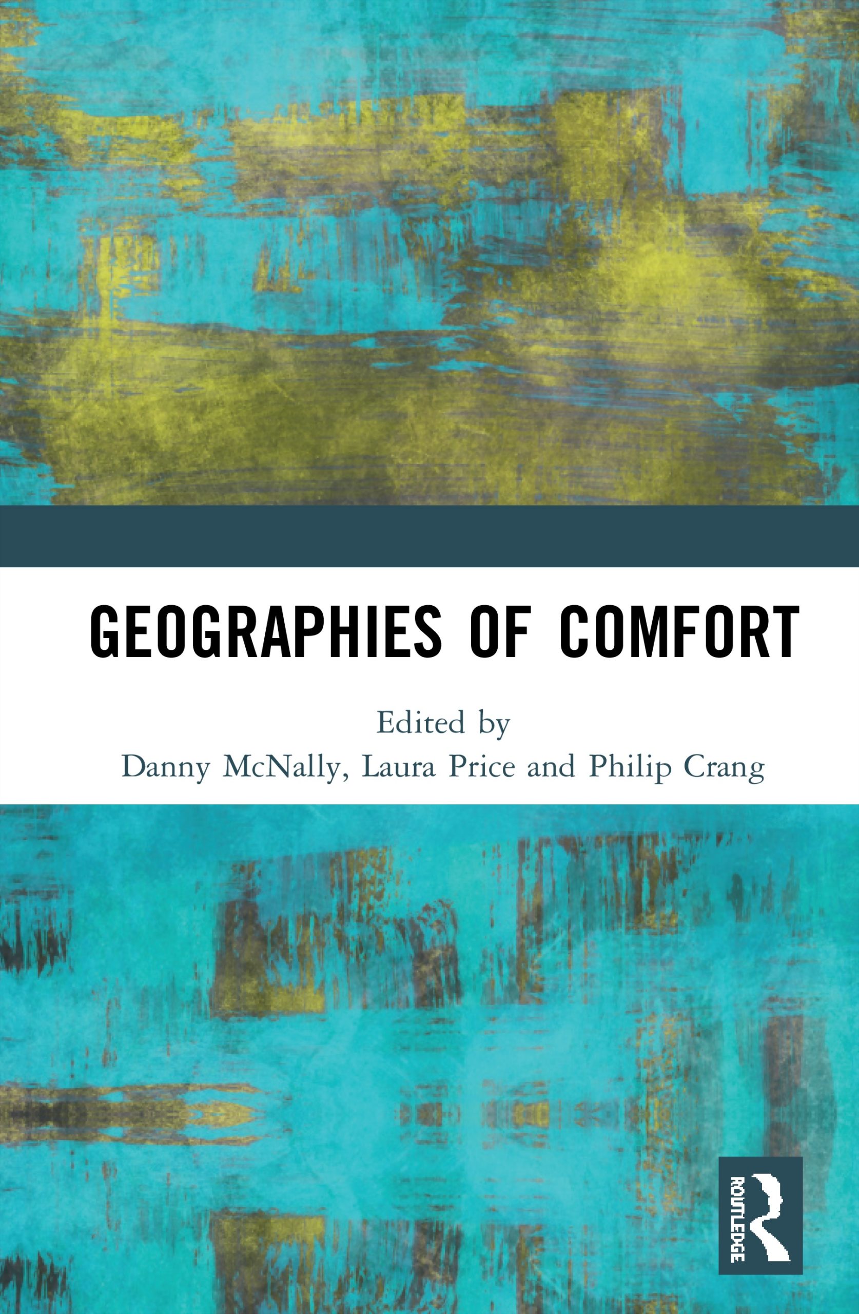
Geographies of Comfort
A volume edited by McNally, Price and Crang.
Bringing together conceptual and empirical research from leading thinkers, this book critically examines ‘comfort’ in everyday life in an era of continually occurring social, political and environmental changes.
Comfort and discomfort have assumed a central position in a range of works examining the relations between place and emotion, the senses, affect and materiality. This book argues that the emergence of this theme reflects how questions of comfort intersect humanistic, cultural-political and materialist registers of understanding the world. It highlights how geographies of comfort becomes a timely concern for Human Geography after its cultural, emotional and affective aspects. More specifically, comfort has become a vital theme for work on mobilities, home, environment and environmentalism, sociability in public space and the body. ‘Comfort’ is recognized as more than just a sensory experience through which we understand the world; its presence, absence and pursuit actively make and un-make the world. In light of this recognition, this book engages deeply with ‘comfort’ as both an analytic approach and an object of analysis.
This book offers international and interdisciplinary perspectives that deploys the lens of comfort to make sense of the textures of everyday life in a variety of geographical contexts. It will appeal to those working in human geography, anthropology, feminist theory, cultural studies and sociology.


